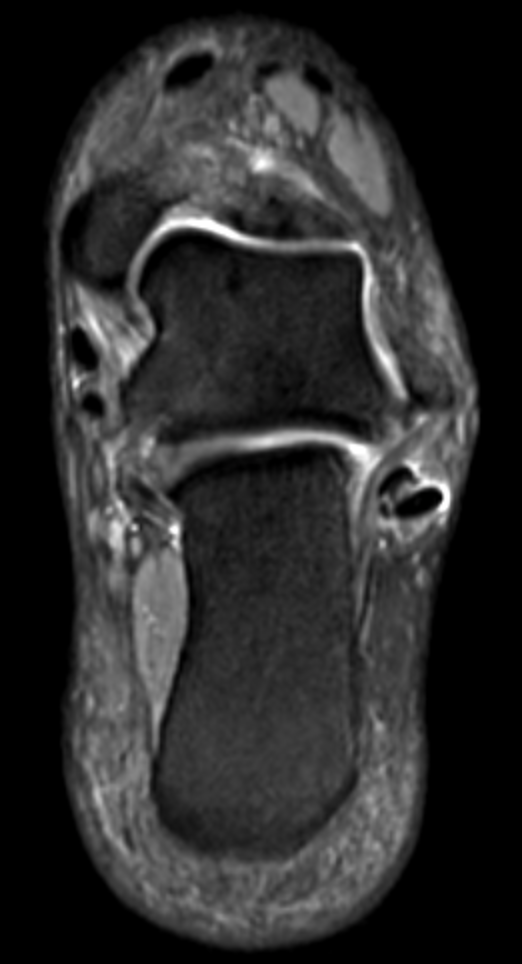
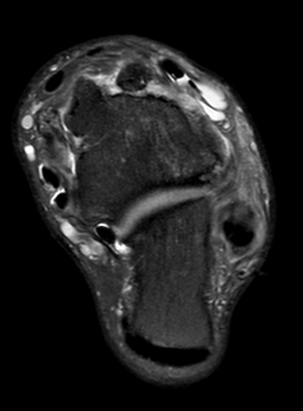
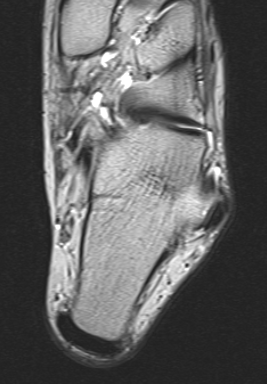
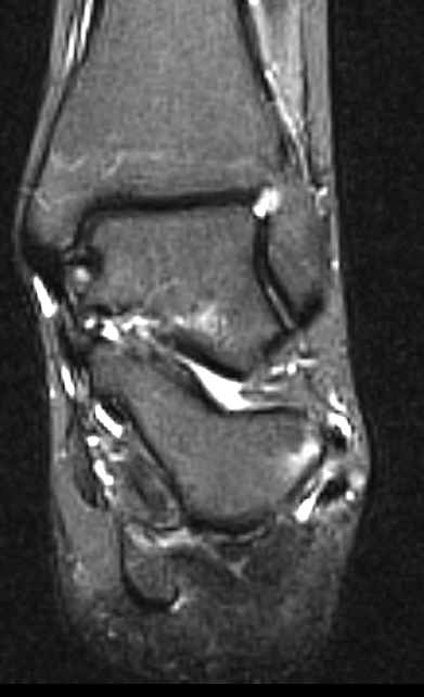
Injuries to the peoneii are most commonly related to misuse.
Peroneus brevis is affected more than peroneus longus
possibly reflecting is position between peroneus longs and the fibula.
The presence of
• an accessory tendon,
• a low musculotendinous junction
• and a prominent peroneal tubercle
may all contribute to the development of peroneal tendinopathy.
A peroneal tubercle elevated more than 5mm from the underlying bone is significant.
The most common manifestation of injuries the development of tendinopathy and longitudinal splits.
Tendon splitting can result in apparent division of peroneus brevis into two tendons which can become displaced to lie in the medial and lateral aspect of peroneus longs.
The identification of three structures within the peroneal sheath should raise suspicion of this injury.
Not all are symptomatic and small splits or defects in the centre of P brevis are seen in asymptomatic individuals
The differential diagnosis of three structures within the tendon sheath includes the presence of an accessory slip of peroneus quadratus.
Peroneus brevis tendinopathy may also occur close to its insertion at the base of the fifth metatarsal.
The most common injury is of course an avulsion fracture called the Jones fracture.
Fractures of an accessory ossicle in peroneus longs may simulate tendon rupture
The peroneal tendons are held in place by and overlaying retinaculum which attaches to the posterolateral margin of the fibula.
Tears of this retinaculum allow the tendons to sublux so that they lie anterior rather than posterior to the fibula.
MOst often this is seen following significant trauma, frequently in association with fracture
More common than this, on dorsiflexion and internal rotation the longus and brevis tendons changing their positions but remaining posterior to the fibula.
This is called internal subluxation and
is a common cause of snapping on the lateral aspect of the fibula.
A peroneus quartus is a common accessory muscle and tendon which may also lie in the peroneal sheath.
The tendon inserts onto the os calcis
Peroneus tertius is another accessory muscle, but this lies anteriiot to the fibula


