Achilles tendinopathy is a common cause of posterior calf pain.
The aetiology is unclear with many factors contributing including overuse and foot mal-alignment, particularly hyper-pronation.
Various manifestations of the disorder are described.
• Tendinosis:enlargement without symptoms
• Tendinopathy: Enlargement with symptoms pain particularly on exertion
• Delaminating tear or delamination
• Partial tear
• Tear
• Paratenonopathy
• Pre and post achilles bursitis
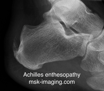
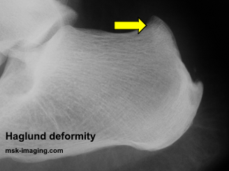
• Enthesopathy Two distinct types are present termed insertional and non-insertional. The commonest of these is non-insertional tendinopathy which typically involves the proximal two thirds of the tendon. Insertional tendinopathy can be due to systemic inflammation esp Reiters syndrome or mechanical eg Haglunds syndrome.
IMAGING
On imaging there is
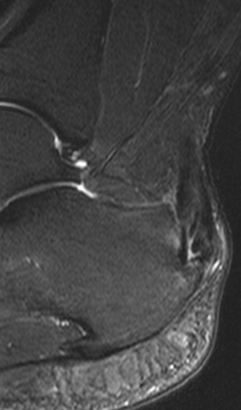
• loss of the normal low signal(mr) of fibrillar(US) internal structure tendon
• enlargement particularly in its AP diameter more than 6 or 7mm.
• Areas of focal or linear increased signal (splits)
• partial tearThere is loss of the normal reflective echo structure. Focal degeneration and partial tears can be identified as areas of focal loss of reflectivity. In addition, ultrasound combined with Doppler imaging demonstrates areas of angioneogenesis. Chronic tendon degeneration perhaps related to hypoxia serve as a stimuli for new vessel formation. New vessels are distributed in a haphazard fashion and are often blind ending. There is some correlation with symptoms probably as a consequence of aberrant nerve growth that accompanies vascular ingrowth. Insertional Achilles tendinopathy involves the distal one third of the tendon. Whilst this may be mechanical in aetiology, sometimes the consequence of bony overgrowth of the posterosuperior margin of the os calcis, a condition termed Haglund’s syndrome, other cases are associated with inflammatory arthropathy.
Rupture of the Achilles tendon is a common event. The condition most commonly occurs in the 30 – 50 age group and is frequently associated with racquet sports such as badminton, squash and tennis. It has been argued that unaccustomed exercise without warming up can predispose but it is likely that there is a significant contribution from underlying Achilles tendinopathy. Patients classically complain of a sudden and acute event where they get a sensation of being kicked in the back of the ankle. An audible popping may also be described. There is a loss of plantar flexion and ability to stand on the toes. Clinically a palpable defect may also be felt. The Achilles tendon tends to rupture in one of three locations. The commonest is in the central third involving the tendinous portion between 4 and 7cm above its insertion. The second commonest is a rupture that occurs at the musculotendinous junction often in or around the level of soleal incorporation. The least common injury occurs with the tendon insertion where an avulsion may occur. Avulsion of the tendon needs to be distinguished from an insufficiency fracture of the os calcis which, as a consequence of tendon retraction, may also similate Achilles rupture.


