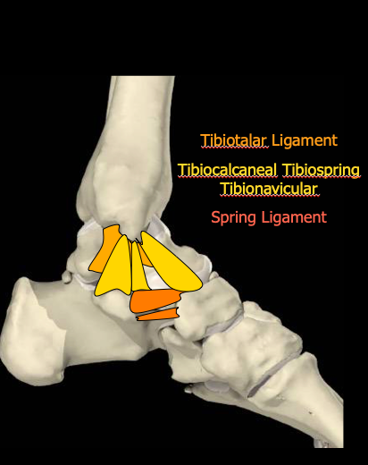
• The medial ligament complex comprises the deep tibiotalar ligament
• which is sometimes divided into the anterior and posterior components
• and three overlying superficial ligaments
• tibionavicular tibiospring and tibiocalcaneal ligaments and
• A transverse component which is the spring ligament
The ligaments on the medial aspect of the foot are less frequently injured than those on the lateral side. The tibiotalar ligament may be injured either by distraction or compression. Compression injury is occur in conjunction with lateral ligament tears.
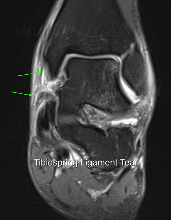
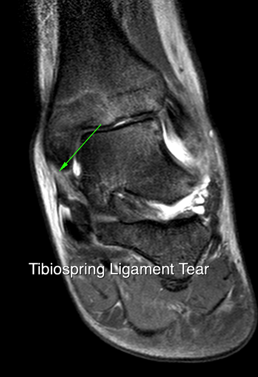
The normal ligament is most reasonably seen on coronal T1 images where the normal striated appearance of connective tissue interspace with fatty tissue is seen. Loss of this normal striated appearance there is a sign of acute injury Chronic changes include loss of the normal striated appearance, calcification/ossification and areas of vascular ingrowth At painful chronic ligament injury is referred to as Posteromedial impingement Ligament avulsion whether it will medial or lateral may be associated with injuries to the periosteal attachments and surrounding retinacula
These include the extensor retinaculum and superior peroneal retinaculum The latter may be resulting in peroneal tendon dislocation.
Injury to the tarsal retinaculum is rare but can result in subluxation of the tibialis posterior tendon
SPRING LIGAMENT
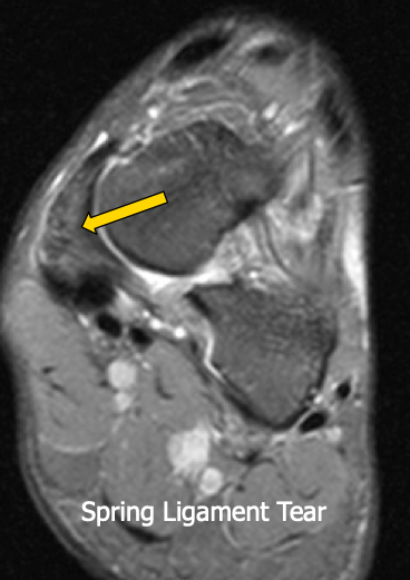
The spring ligament has multiple components . The most relevant for imaging is the superomedial component which is thickened proximally close to its attachment to the sustentaculum and then passes anteriorly and superiorly deep to the tibialis posterior tendon to insert on the navicular. It can be identified either by its relationship to the tibialis posterior tendon or by following the tibial spring ligament on coronal images. There are two further components to the spring ligament which lie more inferiorly and are not thought to tear often Tibialis posterior is separated from the underlying spring ligament by the so called glidinge layer which may be comprises small bursa or thin strip of fibrocartilage.
Injuries to the spring ligament most commonly occur in conjunction with chronic tibialis posterior tendon disease. Failure of the tibialis posterior tendon places a strain on the spring ligament which subsequently fails. Tears that occur as a result of direct injury are less common
but may be the result of landing heavily on the feet and as an overuse injury with similar aetiology to a metatarsal stress fracture.
These injuries tend to occur in younger patients, most commonly males and the injury may follow an unaccustomed increase in impact loading.
Impaction injuries with compression of the ligament against the lateral aspect of the head of the talus have also been described.
ULTRASOUND FINDINGS
Two patterns of ultrasound abnormality have been described. One is complete disruption of a portion of the ligament with a fluid filled gap. The other involves thickening of the ligament with loss of the normal fibrous architecture, decreased signal and increased Doppler activity. The normal size of the ligament is up to 7mm in asymptomatic individuals.
Injuries to other components of the calcaneonavicular ligament are less commonly encountered and are difficult to diagnose. Secondary intertarsal subluxation has been described following complete rupture of the spring ligament. There is plantar rotation of the talus and valgus angulation of the calcaneus. There may be a dorsal subluxation of the navicular. The differential diagnosis of injuries to the spring ligament or talonavicular ligament includes a stress fracture on the dorsal aspect of the navicular. Such fractures occur in the area between the first and second metatarsals related to differential compression forces.


