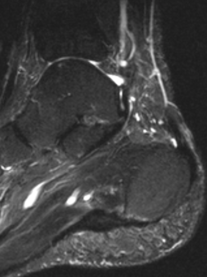
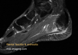
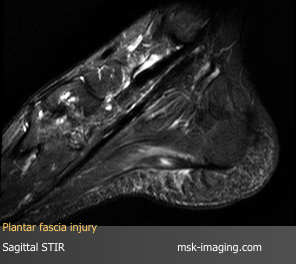
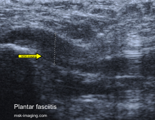
Also called the plantar aponeurosis, the plantar fascia comprises three bundles: central, lateral, and medial.
The medial bundle is the least significant of these and arises from the midportion of the central bundle
The central bundle is the most important and the one most commonly affected by disease.
It runs from the medial tubercle of the os calcis distally blending with the deep fascia and transverse ligaments
The lateral bundle lies beneath the abductor digiti minimi.
The medial calcaneal nerve innervates
Plantar fasciopathy is a common disorder that generally affects individuals in the fourth to fifth decades.
There are several underlying conditions or associations, including an increase in the body mass and
occupations that involve prolonged standing or marching such as in military personnel.
Medical predisposing conditions include diabetes mellitus, seronegative and seropositive arthritides chemotherapy, retroviral, gonococcus and TB infection.
Patients complain of a sharp pain following rest particularly first thing in the morning.
Initially walking helps but symptoms recur with increasing activity.
The area is tender to palpation and there may be decreased dorsiflexion.
Bone spurs are unhelpful as many are incidental.
On X Ray, soft tissue swelling around the spur should be sought and may help identify a symptomatic spur
The plain radiograph should also be scrutinized for other features of enthesopathy at the Achilles tendon insertion.
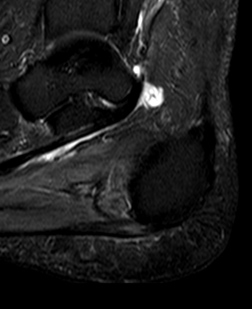
The principal findings on MR imaging in plantar fasciopathy are
diffuse thickening of the fascia with increased signal on T2
surrounding soft tissues on fluid-sensitive sequences.
increased signal at the bony attachment
Coronal MR images are best to differentiate the involvement of the central versus the lateral bundle
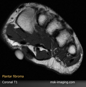
A plantar fibroma is an oval-shaped area of thickening and disorganization intimately relation to the plantar fascia.
Larger lesions may be lobulated and can demonstrate a central scar
A organized internal architecture differentiates from more agressive lesions.
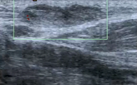
On ultrasound, the lesions demonstrate mixed, predominantly low reflectivity
US is superior to MRI in the detection of lesions that are little bigger than the fascia itself
Occasionally there is increased Doppler signal
Lesions are usually multiple and often bilateral
