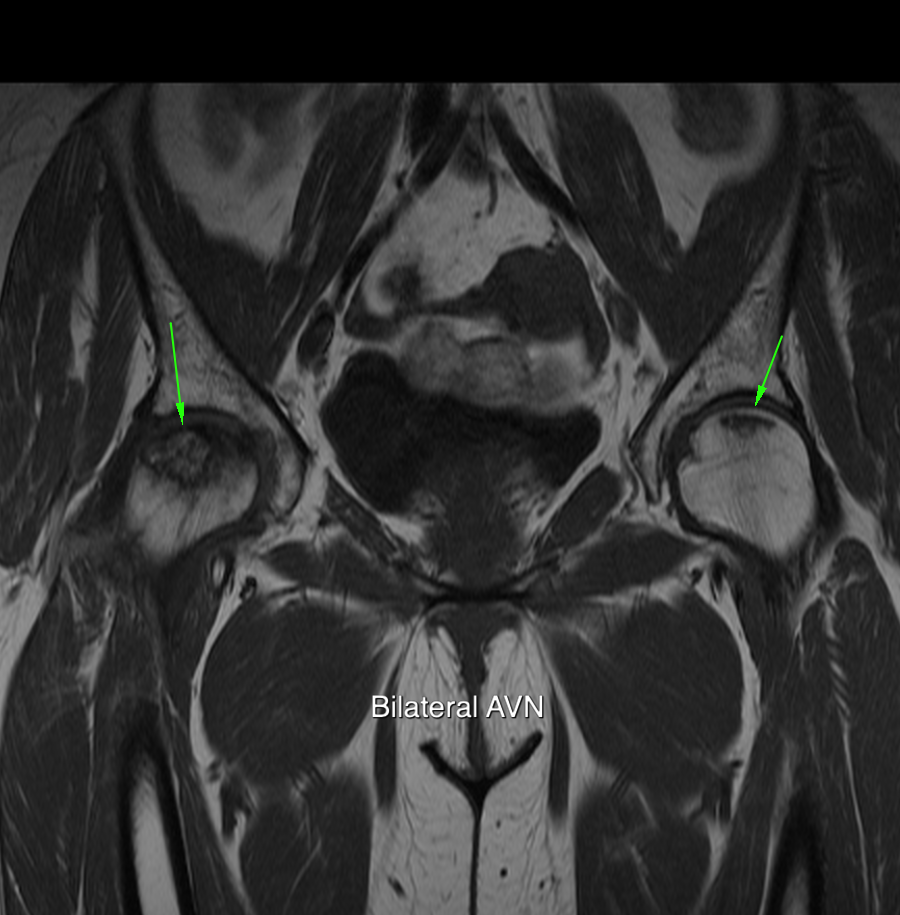
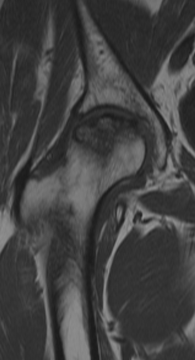
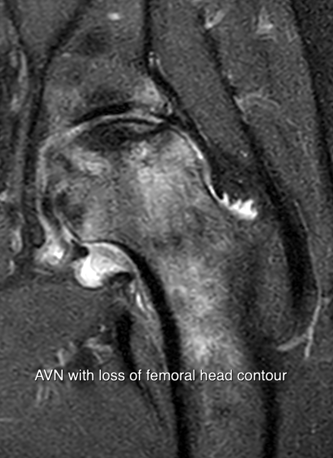
The femoral head is one of the more common locations associated with AVN.
The tenuous vascular supply is prone to interruption
Causes
• Steroid and immunosuppressive therapy
• Alcohol abuse,
• liver and pancreatic disease. R
• vasculicities
• thromboembolic disorders
• sickle cell disease.
• Dysbaric osteonecrosis
The imaging goalsare detection, differential diagnosis and staging.
Detection best with Coronal T1 weighted MR imaging
Both hips should be included
as early asymptomatic disease in the other hip is common.
T1 weighted images are usually supplemented by T2 or FAT saturated images.
Typically AVN lies in the anterosuperior femoral head.
Lesions are variable size
Appearance depending on the stage of the development
most common finding is a well defined geographical area of
loss of T1 signal intensity
without much associated oedema.
and double line sign on T2
Other variants include Fatty type with high signal on T1 presumably representing saponification of the marrow. Fluid and more sclerotic variants occur.
The principal differential diagnoses are subchondral cyst formation secondary to articular cartilage disease and, most importantly, subchondral insufficiency fractures. Avascular necrosis can usually be differentiated from subchondral cyst formation. In avascular necrosis, at least in the early stages, the joint space is preserved and the acetabular side of the joint is not usually involved unless there is secondary degeneration. The so called double line periphery is absent. Avascular necrosis also tends to be anterosuperior within the femoral head whereas subchondral cyst formation related to osteoarthritis is more often a little more superiorly.
The subchondral insufficiency fracture is the most important differential diagnosis of avascular necrosis. The typical imaging finding is a subchondral low signal line with oedema between the line and the joint. This combination can give it a geographical appearance which mimics avascular necrosis. There is a strong suspicion that many cases of avascular necrosis that have resolved may in fact have represented a misdiagnosed subchondral insufficiency fracture in the first place. The differential diagnosis also has important implications for management.


