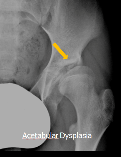
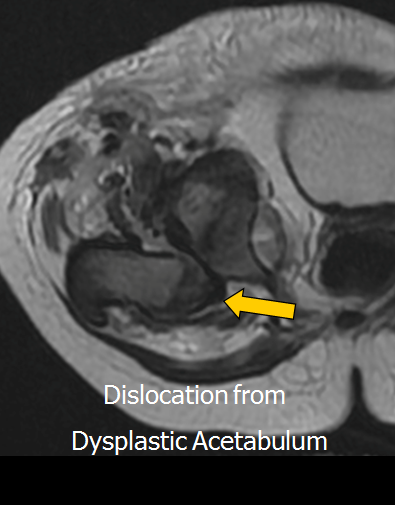
Used to be called congenital hip dislocation.
Change reflects aspects of the aetiology of this condition i.e
• Dysplasia may not be present/obvious at birth and
• The hip can develop normally if treated early.
• Hip dislocation is an extreme component of this syndrome.
Acetabular dysplasia without dislocation or acetabular dysplasia with subluxation are important manifestations of the condition.
Developmental dysplasia is common.
There are some ethnic variations in that the condition is rare in Eskimo’s and Native American Indians.
Once explanation for this is that in these cultures, infants are carried on the mother’s hips for a longer period and that this position abducts the hips and protects against the development of acetabular dysplasia.
Certain conditions predispose to developmental dysplasia including
• a family history
•, a breech presentation,
• spinal, foot and neck anomalies.
Early diagnosis is mandatory to prevent hip subluxation and deterioration of acetabular dysplasia.
Traditionally the diagnosis has been based on clinical examination with the Ortolani and Barlow tests.
Unfortunately these clinical tests will only detect occult hip subluxation and dislocation but
will not detect acetabular dysplasia when the femoral head is completely stable.
Plain radiographs can add to the diagnosis of acetabular dysplasia
however as large components of the femoral head and acetabulum remain unossified the diagnosis can be difficult to establish in the first few months of life.
The iliac angle is most commonly employed
Ultrasound has been used for many years in both Europe and North America to make the diagnosis.
The ability to identify the unossified femoral head cartilage and acetabulum allows a more precise estimation of the relationship between these two structures.
Acetabular dysplasia predominantly affects the anterior acetabular wall and roof leading to reduced containment of the femoral head.
On ultrasound, this can be assessed by calculating the proportion of unossified femoral head that is covered by acetabulum and acetabular labrum.
Two methods have been suggested, one uses two angles (alpha and beta) to express the amount of acetabular development and femoral head containment
the other uses a measurement of the proportion of femoral head covered by acetabulum.
The alpha angle is the angle between the acetabular roof and line drawn along the lateral iliac margin called the iliac base line and the
beta angle is the angle between the tip of the acetabular labrum and the iliac base line.
A decrease in the alpha angle and an increase in the beta angle diagnoses and quantifies acetabular dysplasia.
This approach is used in Austria and Germany
In North America the percentage of femoral head covered by acetabulum is favoured.
A line drawn along the iliac margin is once again taken as the base line.
The proportion of femoral head that lies on the acetabular side of this line defines the percentage of femoral head coverage.
Femoral head coverage less than 45% indicates acetabular dysplasia

