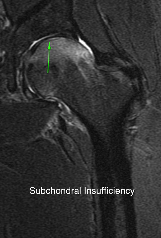
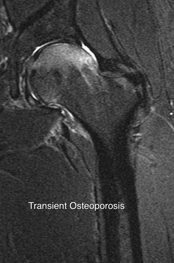
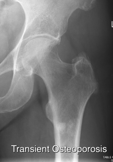
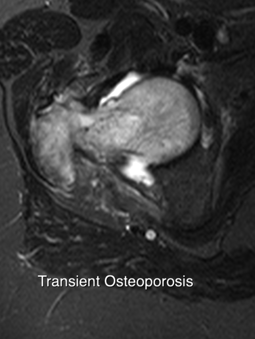
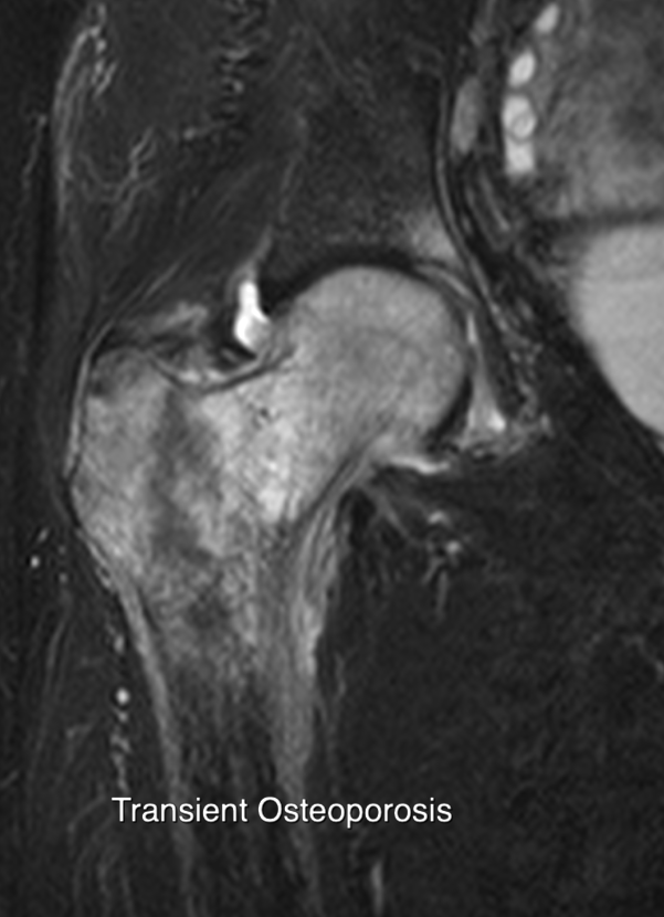
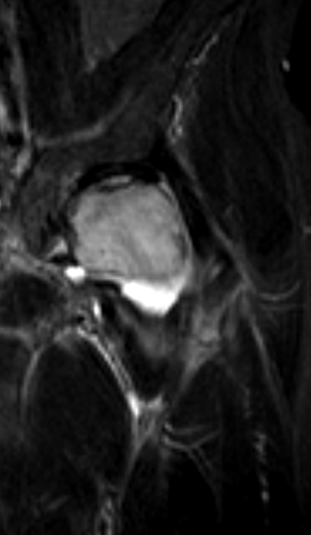
Extensive oedema in the femoral head and neck may be caused by one of the bone marrow oedema syndromes.
These are a group of conditions whose aetiology is poorly understood characterised by
- relatively acute onset hip pain
- associated with osteoporosis on the plain film and
- bone oedema on MR
- involving the femoral head and neck
- extending into the metaphysis
The conditions occur in a number of well defined groups of patients and have several names including transient osteoporosis of the hip, regional migratory osteoporosis and bone marrow oedema syndromes. It may be that time will ultimately show these conditions have distinct features representing different aetiologies however at present they are generally regarded as different patterns of the same disorder.
Transient osteoporosis
most commonly occurs in middle aged men
It is best known however in pregnant women
most common in the third trimester.
Symptomatically patients present with pain and restricted joint movement. The hip is the most frequently involved joint although the talus and knee are also often affected. Any bone may be involved. The left hip is often more involved than the right. The reason for this is not certain but it may reflect the different venous drainage between the two sides and provide some clues to a vascular aetiology. Although one hip may be involved initially, as this improves, the other hip or other joints may become involved. It is these circumstances that the term regional migratory osteoporosis is most commonly applied
The MR imaging features extensive bone oedema
The classic distribution is patchy.
The oedema may have well demarcated areas of involvement with other areas spared (see the figures associated with this description). An effusion, possibly associated with synovitis is common. Occasional the acetabulum may be involved, the precise reasons for this is unknown but it may reflect the associated synovitis. Prognosis The typical pattern is that pain may persist for 6 to 12 months before regressing. As previously outlined regression may occur in one joint and then recur in another. MRI may remain abnormal for some period beyond this. There is good evidence to suggest that there is a background bony anomaly and that transient osteoporosis represents a more diffuse background osteoporosis. It is therefore worthwhile carrying out DEXA in these patients to detect such an anomaly.
