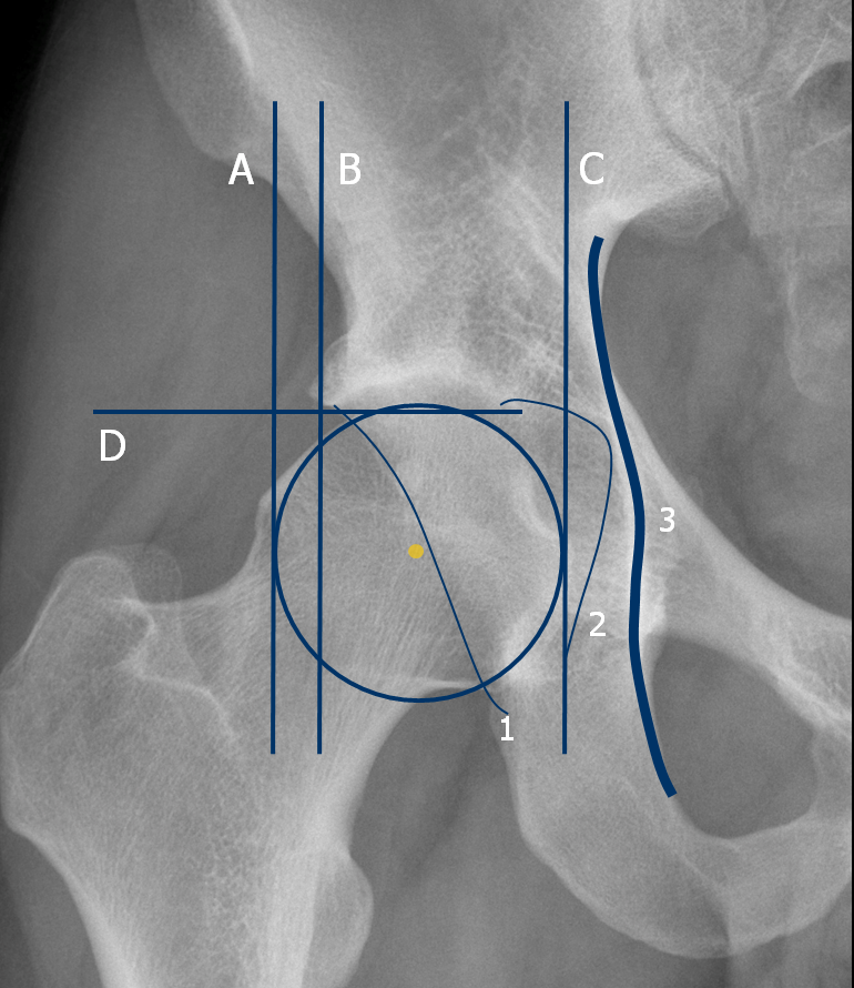
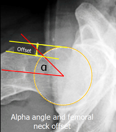
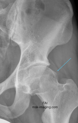
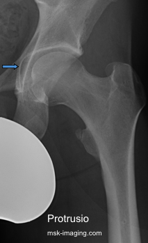
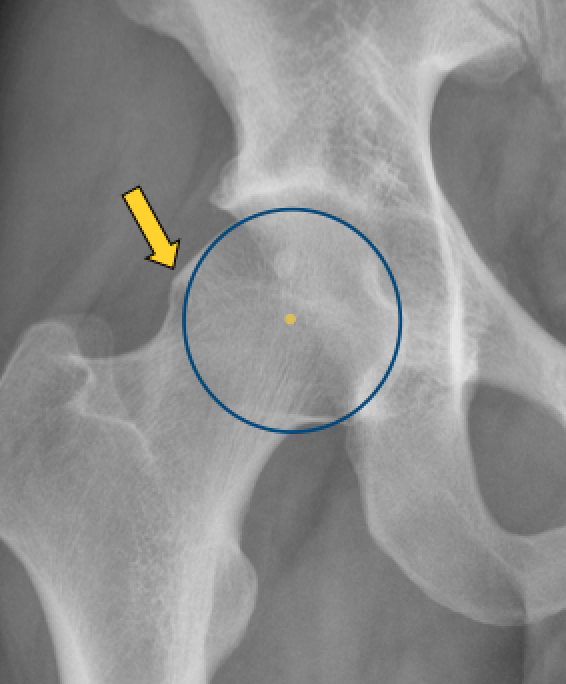
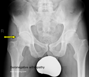
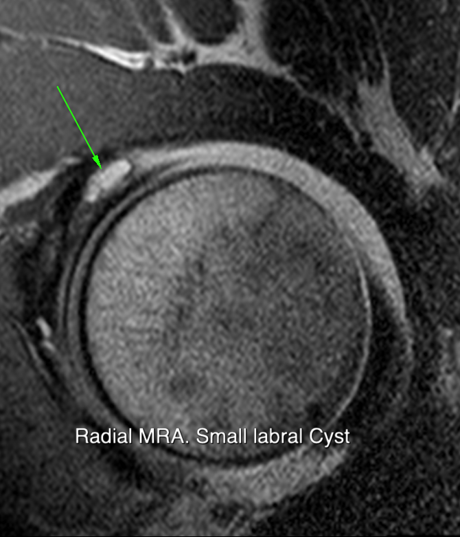
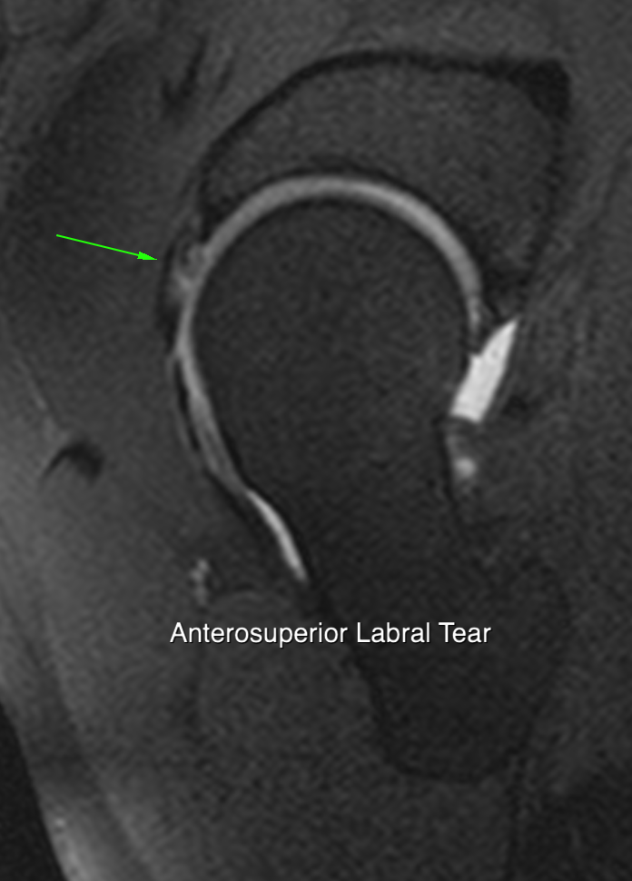
There are a large number of measurements that the X Ray report could include to describe the shape of the proximal femur and its relationship with the acetabulum. Careful attention to technique is needed to ensure standardisation before ascribing significance to any measurement. Some possible measurements include:
1 Cam lesion (AP & shoot through lateral)
• a Subjective: presence and rough estimate of size
• b Alpha angle on lateral (image 3)
• c Anterior offset on lateral (image 3)
2 Anterior cortical reactions or herniation pits
3 Acetabular protrusion (when line 2 crosses line 3 by >3mm in males 6mm in females)
4 Centre edge angle (image 1- measurement of dysplasia)
5 Acetabular Index (image 1- line joint margins of sourcil-overcoverage when less than 0 degrees)
6 Extrusion index (ab/ac on image 2)
7 Sharp angle (angle of acetabular abduction)
8 Anterior centre edge angle (from lateral xray)
9 Acetabular wall relationship to femoral head (line 1 and dot on image 2)
10 Crossover sign (image 4 - ant wall does not normally cross posterior wall)
11 Pelvic inclination - from lateral
1 & 2 measure cam and its consequences
3-10 measure acetabular dyplasia and coverage
The cam lesion is seen as infilling between the femoral head and neck. On plain radiographs, it is detected using an anteroposterior and a cross table lateral view of the proximal femur. On the AP view, the best visualisation of the head neck junction is obtained with the images obtained with the leg in 15 degrees of internal rotation. The beam should be centred at the mid point of a line joining the anterior superior iliac spines directly superior to the pubic symphysis. An assessment of pelvic tilt on the lateral radiograph can also be made to determine whether the radiograph can be reliably used in the assessment of FAI.
Pincer impingement or acetabular over coverage is more common in females. Over coverage can be general or focal, depending on the abnormal anatomy, either
• anterosuperior overhang
• coxa profunda & acetabular protrusio or
• acetabular retroversion.
General over coverage occurs with Cox's profunda and protrusio acetabuli. Coxa profunda is diagnosed when the acetabular fossa line touches the ilioischial line. Protrusio acetabuli is a more advanced form of this, when the femoral head overlaps the ilioischial line.
Focal over coverage can occur either anteriorly or posteriorly. Anterior over coverage occurs with acetabular retroversion and posterior over coverage when there is a prominent posterior wall.
A number of other measurements may be associated with acetabular over coverage. The posterior wall sign where the wall of acetabulum lies medial to center for femoral head is very suggestive of acetabular retroversion.There may be an increase in the centre edge angle and a decrease in the acetabular roof index. The proportion of femoral head coverage decreases and the posterior acetabular wall projects over the centre of the femoral head.
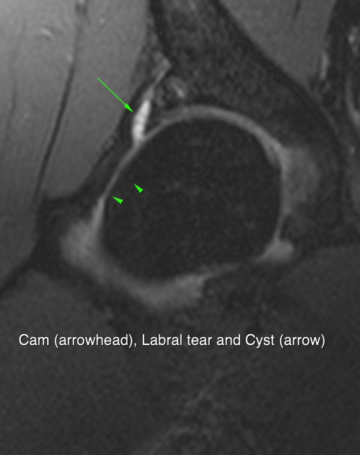
Presence or absence of cam lesion
Extent of cam ie how many slices are abnormal (Omega angle)
Cam can also me measured as alpha angle (like the xray alpha angle)
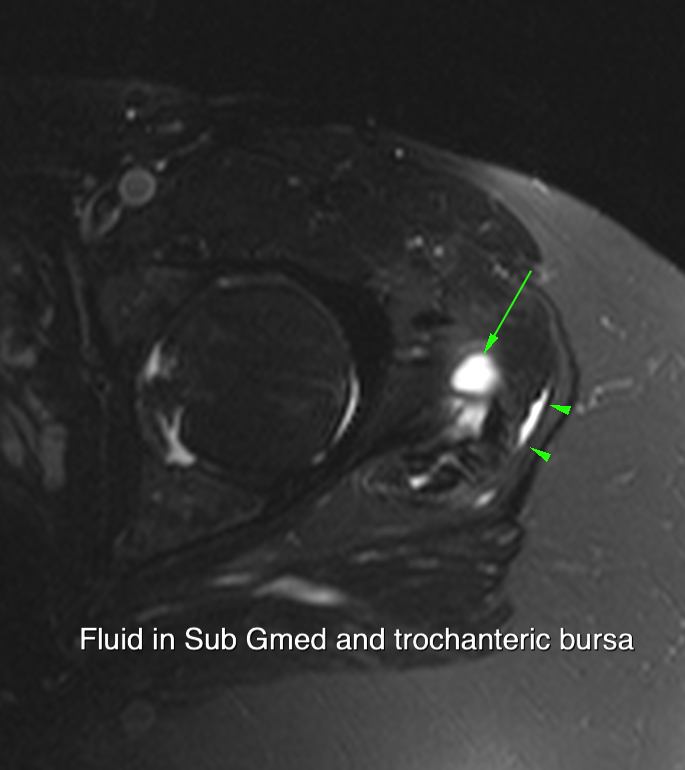
Status of labrum
Presence of cartilage lesions
Don’t forget the posterior joint space and posterior labrum
See next section

