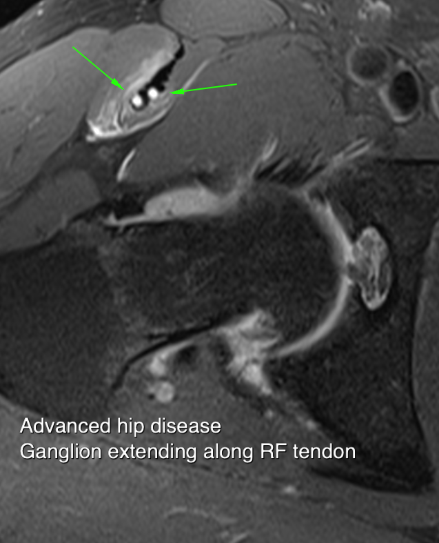
The differential on sport related groin pain is wide and includes:
- Rectus abdominis tears or haematoma
- Symphysitis
- Adductor origin/insertion enthesopathy
- Direct and Indirect inguinal hernia
- Femoral hernia
- Hip disease
- rectus femoris tears Sartorious origin injuries.
True hernias are rare in young patients. A common dilemma is to distinguish adductor disease from the so called sportsman's hernia. Males are more often affected and pain at the end of the game is a common complaint. .
IMAGING
Sagittal images provide excellent visualisation of the condensed insertions off rectus abdominis and the adductor longus. The inguinal ligament and conjoined tendon insert from the lateral side.These are supported by oblique axial plane images which show the pubic symphysis to good effect. Typical findings include bone oedema in the pubic synthesis and surrounding adductor tendon.A cleft may extend from the symphysis and pass under the adductor origin as the tendon is lifted off bone.Occasionally gadolinium enhancement is useful in making this assessment
INGUINAL HERNIA
The important locations for hernia in the patient with groin pain are the inguinal and femoral canals. Within the inguinal canal, there are two types of hernia, indirect and direct. An indirect hernia is one that travels along the length of the inguinal canal. and indirect hernia enters the canal via the deep ring and travelled medially along the canal itself emerging, it's long enough, from the external ring. A direct hernia is one that enters the canal through a week or defective posteriorly and then travels along the canal either in the medial or lateral direction. The particular characteristics of an indirect inguinal hernia are therefore enters via the internal ring begins lateral to the epigastric artery travel is parallel to the inguinal cana At rest, hernias can be either reducible or irreducible. And irreducible hernia is one that is present within the Kanow at all times. Reducible hernia, the more common in younger individuals, is one that is only present when there is an increasing abdominal pressure. Dynamic imaging is therefore required to diagnose this common type. The dynamic imaging used is most often ultrasound. Dynamic MR imaging during Valsalva manoeuvre can also be used but it is more difficult to produce a reliable Valsalva using MR. The ultrasonologist however can feel whether the patient is performing the manoeuvre correctly and therefore ultrasound provides a more reliable true negative test The ultrasound technique therefore is to locate the internal ring and inguinal canal with the patient relaxed. The patient is then encouraged to perform some form of strain manoeuver. The competency of this is best assessed without ultrasound probe in hand and the patient should be encouraged to practice until there is clear cut evidence that a high pressure strain is being performed. Once the dynamic manoeuve has been successfully taught, the probe is placed parallel to the inguinal canal. If an indirect hernia is present, a mass seem to enter via the internal ring and travel along the canal. The ma bowel but more often comprises omental fat. ....
ADDUCTOR INJURIES
Patients with groin pain can be broadly divided into two groups.
Those were the pain emmanates from at and above or below the level of the pubic symphysis
In the case of the latter group, the adductor origin represents a significant cause. The adductor longus muscle has two components. A tenderness component which attaches close to the pubic and plans with the fibrous upon your roses that covers the synthesis and blend superiorly with the rectus abdominis tendon. The muscular component extends immediately lateral from the fibrous attachment and extends the muscle origin alarm the inferior pubic ramus. The commonest location for injury is that the tendon attachment itself.
Injuries can be acute or chronic, all the more likely the latter is acute on chronic.
The spectrum of abnormalities present in this location include
- adductor tendon avulsion
- chronic adductor tendinopathy
- adductor cleft
- adductor muscle tear
- osteitis pubis


