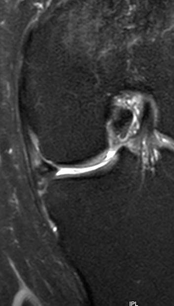
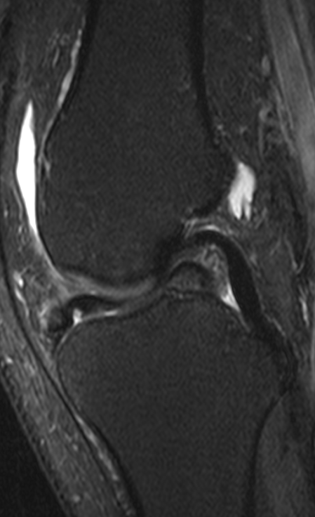
Tear where a significant portion of the meniscus becomes separated from the underlying meniscus and displaces .
Ddisplaced fragment usually large.
Look for :
• reductionin the size of the meniscus
• Filling defect in the notch.
A variant is where the fragment displaces anteriorly rather than centrally.
MR demonstrates apparent enlargement of the anterior horn compared with the posterior horn.
This change in morphology is key to diagnosis
Enlargement of the anterior horn has also been called pseudo-hypertrophy..
The most difficult lesions to diagnose are those that involve a small proportion of the meniscus.
These lesions usually start out as simple tears which extend.
The fragment of the meniscus can be displaced:
• externally around the rim of the femur
• around the rim of the tibia
• either superiorly or inferiorly
• posterocentrally into the notch
• or can be pushed along the meniscus creating a raised portion.
An inportant clue is to look for any change in the size or shape of the meniscus including a blunted free edge. Then look carefully in the above places.
Most flaps maintain a tenuous connection with the parent meniscus


