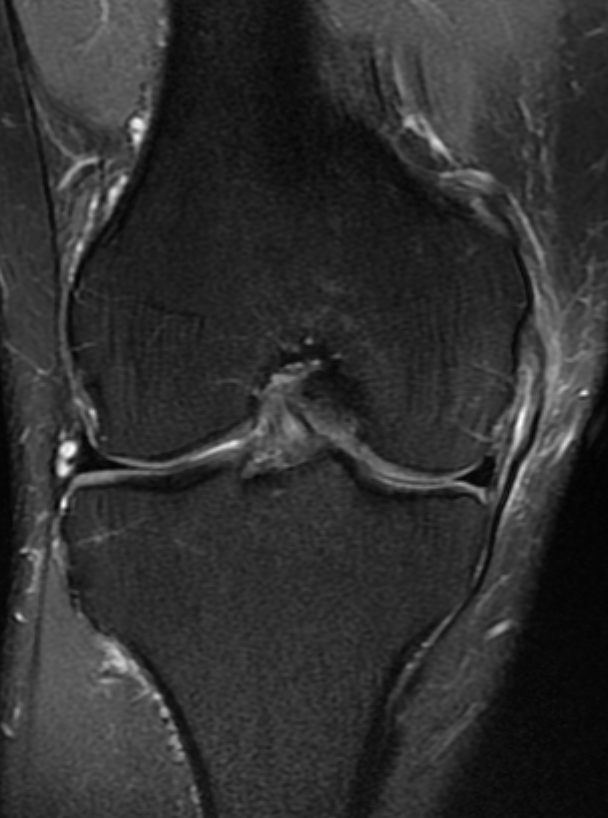
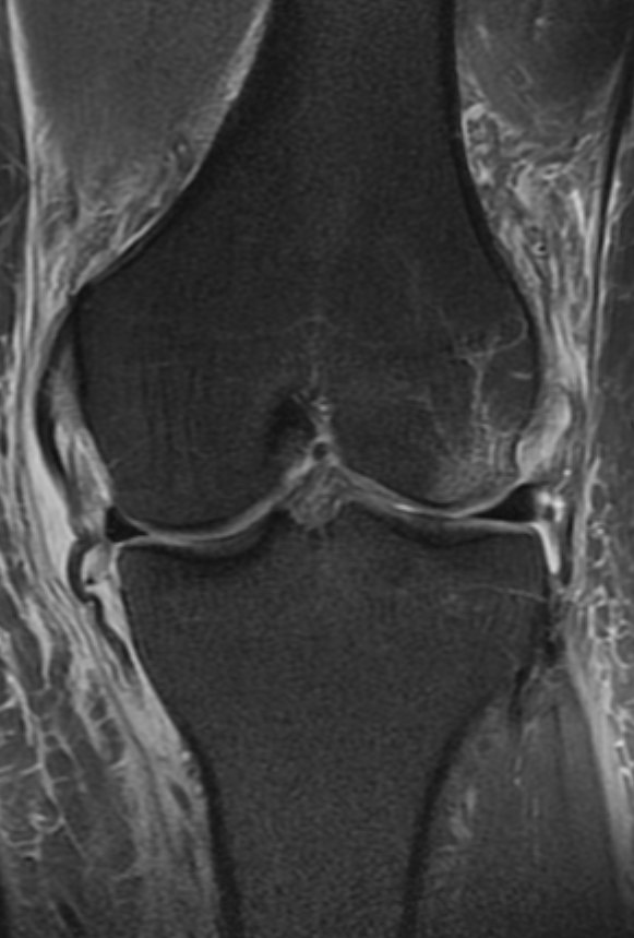
The medial collateral ligament comprises two portions.
The superficial component is stronger
It runs from several centimetres above the knee joint to insert 5-6cm below.
It is approximately 2.5cm anterior to posterior.
Anteriorly it is related to the medial retinaculum and posterior to the capsule of the knee joint
along with as specific condensation that runs posteriorly and obliquely to reinforce the posteromedial aspect
The deep portion of the medial collateral ligament is composed of two weak ligaments, meniscofemoral & meniscotibial ligaments
Between the two components lies the tibial collateral ligament bursa.
The MCL is injured under two circumstances.
The commonest is a valgus sprain which is associated with compression micro fracture in the lateral compartment.
A second mechanism is less common where there is valgus translation of the proximal tibia. This is generally not associated with impaction micro fracture however medial meniscal extrusion may be observed.
Injuries are most easily assessed on coronal images.
INJURY GRADE
• 1 Oedema surrounding an otherwise intact ligament.
• 2 Tear but not complete disruption of the ligament
• 3. Complete disruption.
MCL tears may be associated with pseudolocking.
Grade 2 may have a lamellated appearance described as onion skinning
In most cases the ligament injury will heal without complication although ligament thickening may persist for considerable periods.
The pes anserine tendons comprise the sartorius, gracilis and semitendinosus tendons
Their combined insertion resembles a gooses foot leading to the unusual name.
Injuries to the per anserine insertion may be acute or chronic.
Acute injuries usually result in tendon avulsion.
Chronic injury manifests as per anserine bursitis.
The detection of per anserine bursitis should prompt a search for other potential enthesopathy and bursitis. of the joint along with the insertion of the semimembranosus tendon called the posterior oblique ligament (POL).

