Look for and report
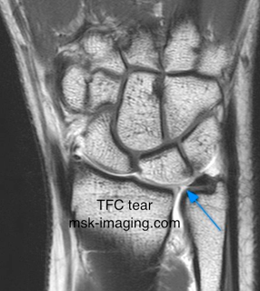
• Defects in the TFC itself
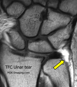
• Tears of the two main ulnar attachments
• Tears of the volar and dorsal ulnotriquetral ligaments
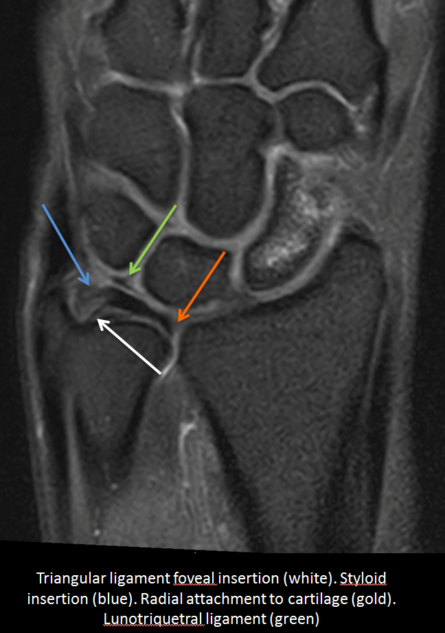
• Lunotriquetral ligament tear
Atzei classification of TFC injury
• 0 Ulnar styloid fracture
• 1 Tear of ulnar attachment - distal (styloid) branch
• 2 Tear of ulnar attachment - both branches
• 3 Tear of ulnar attachment - proximal (fossa) branch
• 3a 3 plus styloid fracture
• 4 Central TFC and both ulnar attachments
• 5 Radial attachment
The TFCC is attached on the radial side to the most distal ulnar part of the radius.
The attachment is not to bone but to a thin layer of articular cartilage
The signal change that occurs at this attachment should not be misinterpreted as a tear.
On the ulnar side, the attachment of the TFC is more complex.
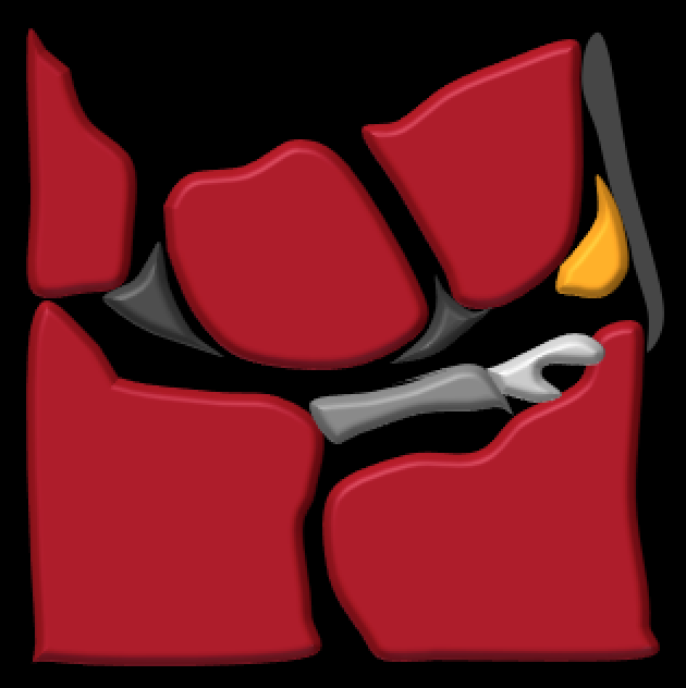
The principal attachment is to the base of the ulnar styloid through a loose ligamentous attachment interspaced with fibre adipose tissue.
See diagram at base of page
There are ulnolunate and ulnotriquetral ligaments on the volar aspect of the TFC
These lie deep to the ulnar nerve - see diagram at bottom of the page
Perforations of the TFC occur with increasing age
Particularly are common over the age of 40.
Clinical correlation thus important
In symptomatic patients, arthographic studies have demonstrated similar findings in the contralateral asymptomatic wrist
Tears of the TFC may be partial or complete.
If partial may involve the proximal (most commonly) or distal articular surfaces
May be on the radial or ulnar sides.
Much needs to be clarified about the clinical significance of these injuries
Currently thought that ulnar sided injuries are more important than radial and
Proximal partial tears are more common, and possibly more clinically significant, than distal partial tears.
Partial tears of the distal surface of the TFC will not be detected by distal radio ulnar joint injection and
Partial tears of the proximal surface will not be detected by the more commonly performed radio carpal injection.
Injection of both compartments may also be required to demonstrate the (rare) unidirectional tear.


