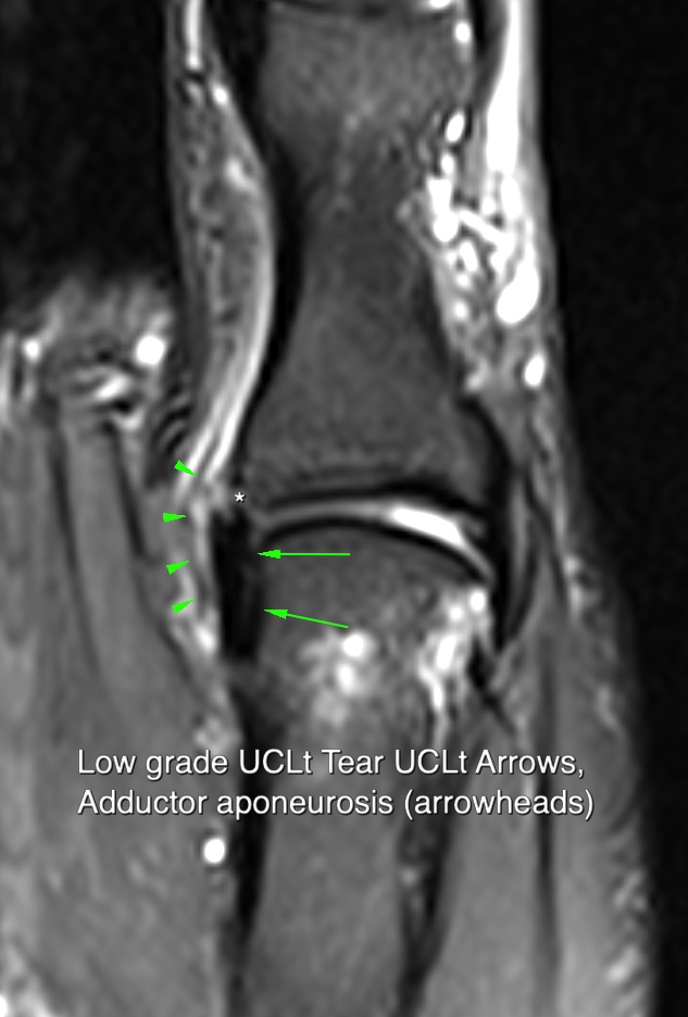
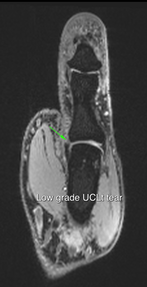
Injuries to the ulnar collateral ligament of the thumb were classically described in game keepers where the injury occurs as a consequence of the tortional movement used to break the neck of rabbits. More commonly nowadays they are seen as a ski-ing injury when the strap on the ski pole forcibly abducts the thumb during a fall. The injury is an avulsion of the distal attachment of the ligament at its insertion into the base of the proximal phalanx.
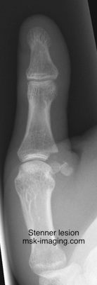
In most cases the avulsion includes a small fragment of bone, in which case the extent of the injury can be determined from plain films. If the tear is minimally displaced, the small bony fragment lies close to its normal location in the base of the proximal phalanx. With more severe injury, at the time of maximal abduction the torn ligament may displace proximal to the palmer aponeurosis. As the joint is reduced the ligament is unable to return to its normal location due to interposition of the aponeurosis. In these instances, the joint remains unstable and healing is impossible without surgical reduction. Displacement of the torn ligament is referred to as a Stenner lesion. Both MR and ultrasound have been used to diagnose a Stenner lesion. The ultrasound technique involves placing the probe on the ulnar aspect of the abducted thumb. The probe is held in the left hand and the examiners right finger and thumb can be used to stress the joint. This can be helpful to determine both the integrity of the ligament and the degree of instability that has occurred as a result of injury. Gentle flexion of the IPJ help to identify the aponeurosis. With both imaging techniques the ligament can be identified displaced and coiled proximally outside the aponeurosis. The ultrasound appearance of the displaced ligament has been likened to a yo-yo.


