Anterior dislocation most common
Heavy fall on shoulder esp in contact sport typical
I Traumatic structural: a) acute b) persistent c) recurrent
II Atraumatic structural a) recurrent
III Habitual non-structural a) recurrent b) persistent.
Surgical treatment involves capsular tightening procedure which can be open or arthroscopic.
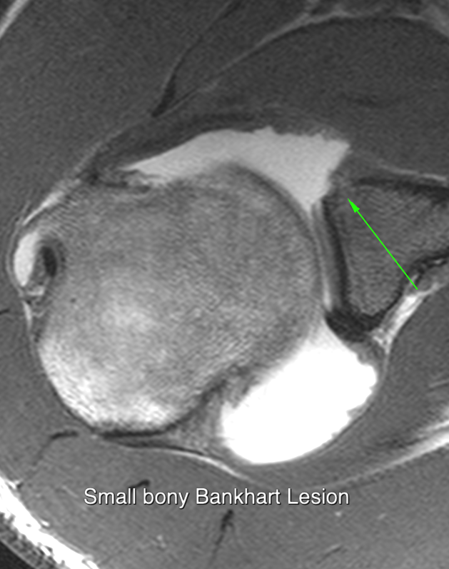
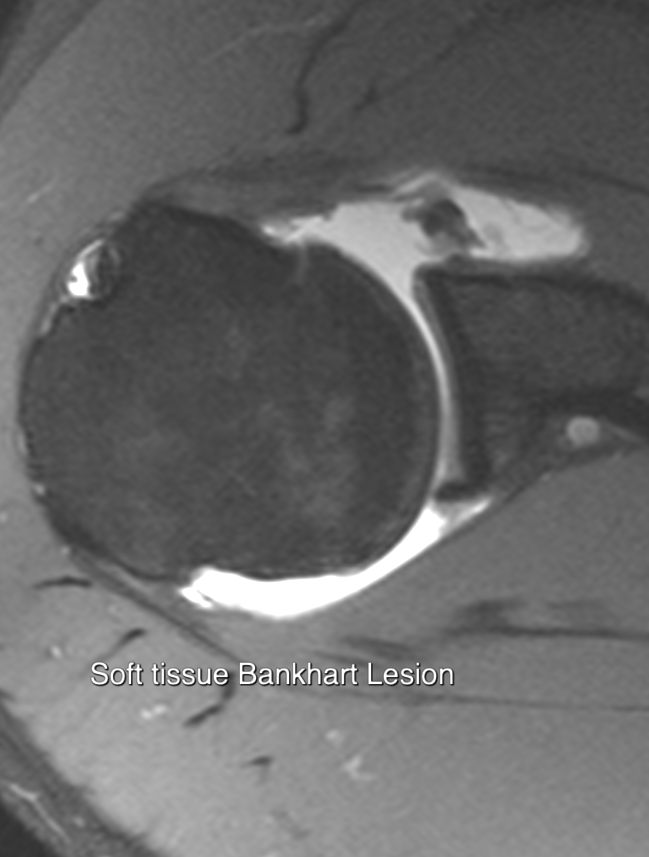
Imaging may be requested to determine whether surgery to the anterior glenoid margin is necessary. Surgery is indicated where there is a significant bony injury (bony Bankart lesion) and in many cases where the injury is confined to the anteroinferior labrum (soft tissue Bankart lesion).
Clinical questions for imaging:
Is there a Bankhart lesion and is it bony or soft tissue
Is there a Hill Sacks lesion and is it big enough to engage
Humeral head is subcoracoid on frontal image when dislocated anteriorly
Posterior dislocation more difficult and needs lateral view
Look for Bankhart fracture post reduction but difficult
Report should comment:
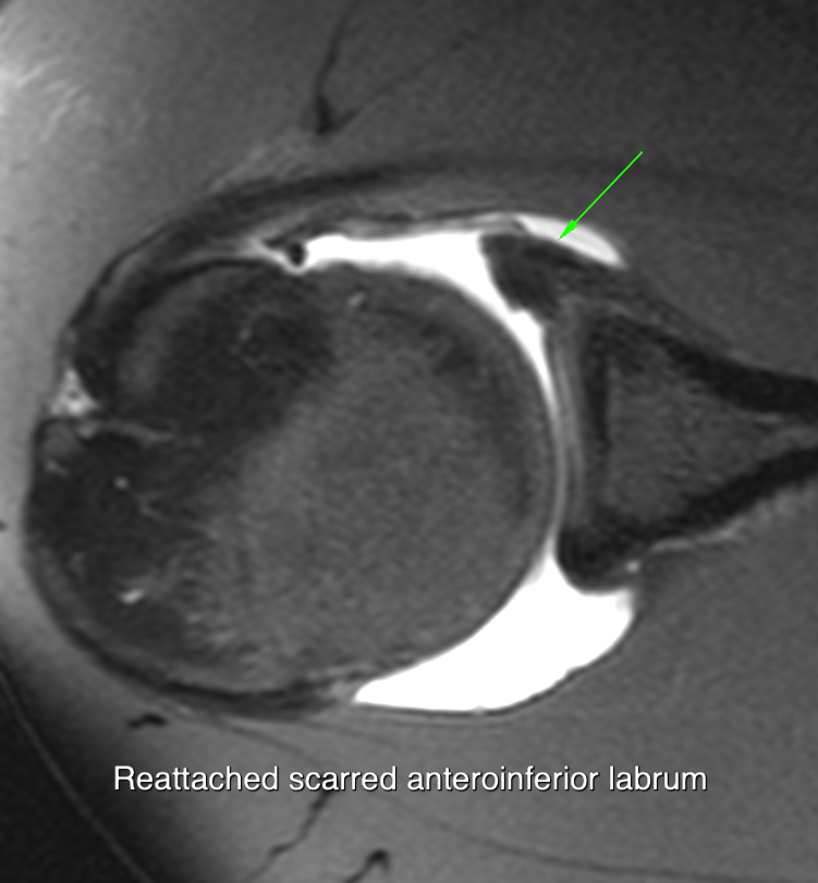
State of anteroinferior labrum and adjacent glenoid
Presence of Hill Sachs lesion and whether it is on track
State of glenoid cartilage
Status of glenohumeral ligaments and capsule
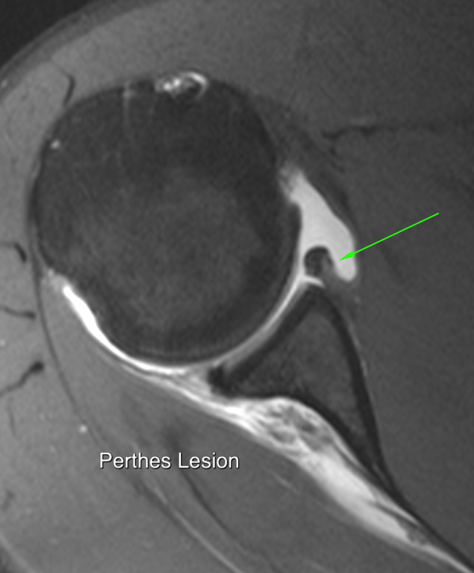
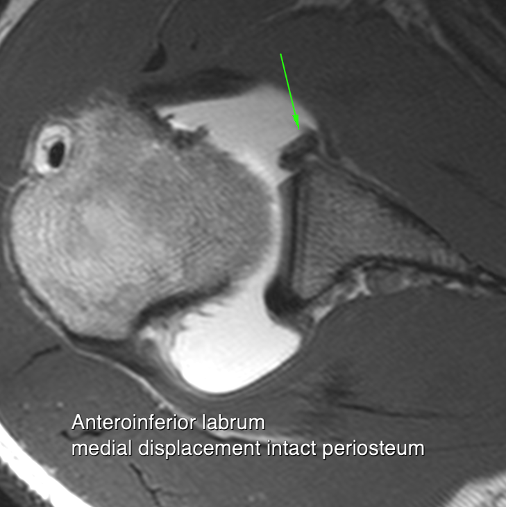
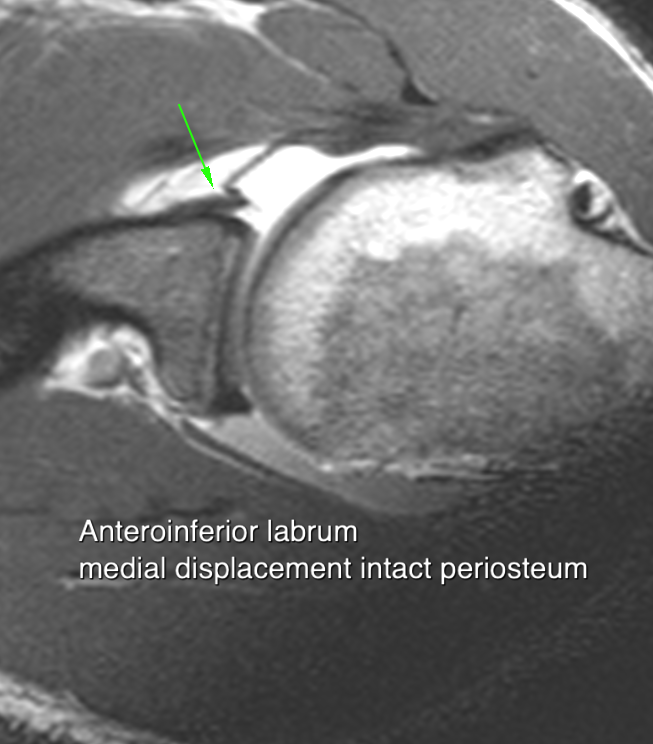
Typically the anteroferior labrum with attached anterior limb of the inferior glenohumeral ligament (IGL) is pulled from its insertion and displaced laterally.
PERTHES LESION Periosteum remains attached to the anterior margin of the labrum
GLAD LESION a small fragment of articular cartilage is attached the labrum
ALPSA LESION labrum is displaced medially(anterior labrum periosteal sleeve avulsion)
GLOM LESION blood supply to the labrum is preserved it enlarges forming a glenoid labrum ovoid mass.
HAGL Humeral avulsion of the inferior GH ligament
The clinical significance of these injury variants is extremely doubtful
A Hill Sachs lesion is an impaction fracture of the posterior aspect of the humeral head
Look for flattening above coracoid level.
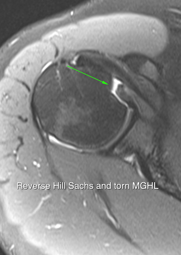
Reverse Hill Sachs lesion occurs with posterior dislocation
Large HS lesions can become engaged on the anterior glenoid during external rotation. In these instances, the surgeon may choose to tighten the anterior capsule to a degree that reduces the likelihood of this engagement occurring.
The track refers to the component of the humeral head surface that articulates with the glenoid. If the Hill Sachs is larger than the width of the track, engagement may occur and this is called an 'OFF TRACK' lesion
The width of the track is 0.83 times the glenoid width, minus the width of any bony bankhart. If the width of the HS lesion is larger than this it is deemed Off Track.
GT = Glenoid Track=0.83* Diameter of the inferior glenoid
EGT=Effective Glenoid track= GT - width of the anterior glenoid bone defect)
HSI = Hill Sachs Interval= width of HS lesion plus width of bone gap between cuff footprint and HS lesion
OFF TRACK if HSI > EGT
The main glenohumeral ligament tear involves the inferior glenohumeral ligament which is divided into anterior and posterior components.
This attachment is best appreciated on MR imaging when the patient is examined in the so called ABER position. This is the position of abduction and external rotation whereupon the patient places his hand behind his head. Middle glenohumeral ligament tears also occur
Almost no role
Anteroinferior labrum can be visualised with 'hand behind head' position
US mainly used to detect associated acute cuff tears


